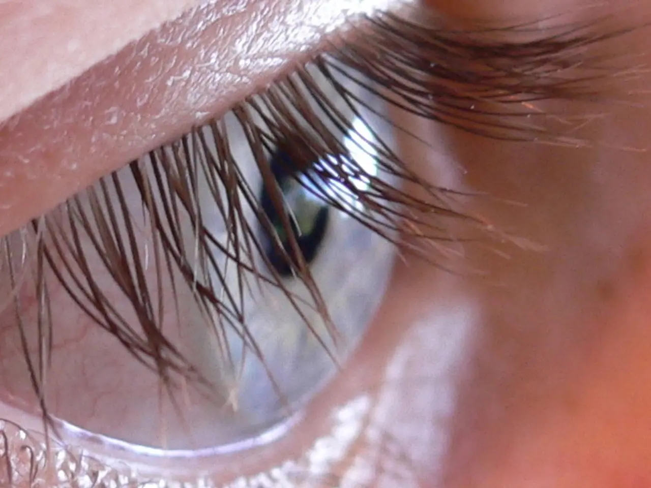
New Imaging Method Reveals Real-Time Immune Cell Activity in Eye
Researchers have made a significant breakthrough in eye health with a new imaging method that allows real-time observation of immune cells in the eye. This innovation, developed in collaboration with institutions from Japan and the US, could revolutionize the diagnosis and treatment of diabetic retinopathy and other retinal diseases.
The new method combines a head-fixation device, contact lenses, and a custom objective lens to provide high-resolution visualization of living retinal cells. In diabetic mice, it was discovered that microglia, immune cells in the retina, become hyperactive before visible vascular damage occurs. This finding suggests that inflammation precedes blood vessel damage in diabetic retinopathy.
The research team, funded by organizations including the Japan Agency for Medical Research and Development and Novo Nordisk Pharma Ltd., also found that Liraglutide, a diabetes drug, normalizes the overactivity of microglia in diabetic mice. Remarkably, this effect is not dependent on blood sugar levels.
The new imaging method, which enables lifelong observation of immune cells in the eye, could lead to earlier detection and more targeted treatment of diabetic retinopathy. Its potential extends to other retinal diseases like glaucoma and age-related macular degeneration. The discovery that inflammation precedes blood vessel damage and the potential of Liraglutide to normalize microglia activity offer promising avenues for further research and treatment.

