
Procedural application of highly targeted artery blockage through a catheter for addressing abdominal lung growths unconnected to the lung
News Article: Minimally Invasive Treatment for Rare Neonatal Condition
Intra-abdominal extralobar pulmonary sequestration (EPS) is a rare congenital anomaly that is typically diagnosed prenatally or in the neonatal period. This condition, which involves a mass of non-functioning lung parenchyma with an anomalous systemic arterial supply, can cause complications such as infection, hemorrhage, or mass effect.
A recent case report involves a 30-day-old newborn who was diagnosed with intra-abdominal EPS associated with left diaphragmatic agenesis. The diagnosis was confirmed through postnatal imaging, including a CT scan, which revealed the non-functioning lung tissue mass with anomalous systemic arterial supply and venous drainage separate from the normal pulmonary circulation.
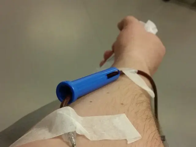
In this particular case, the patient underwent a minimally invasive treatment known as superselective transcatheter arterial embolization. This procedure aimed to occlude the aberrant systemic artery supplying the sequestration, potentially avoiding surgery. The embolization was performed using 0.018" Interlock coils. The accessory lung was devascularized during the embolization procedure.
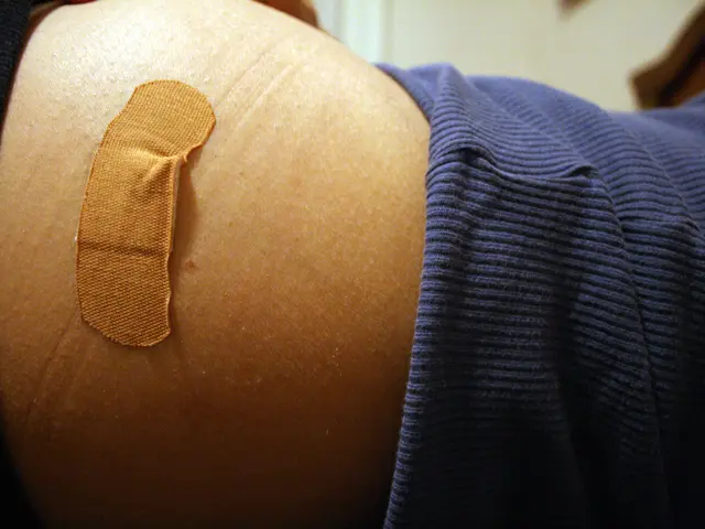
Following the embolization, the patient underwent left thoracotomy with excision of the accessory lung and reconstruction of the diaphragm using bovine pericardium. The patient was discharged from the hospital after 4 days without complications and has remained asymptomatic at 8 months of follow-up, showing appropriate development for his age.
While surgical excision remains the standard treatment for EPS, including intra-abdominal locations, less invasive alternatives like embolization are emerging as potential options in certain cases. These alternatives aim to reduce the invasiveness of treatment and potentially avoid surgery in select cases or be used adjunctively.
It is important to note that EPS is typically diagnosed within the first few months of life. Early detection and treatment are crucial to prevent complications and ensure the best possible outcome for the patient. Prenatal ultrasound, particularly detailed sonography, can detect intra-abdominal EPS as a mass, often suspected due to its characteristic systemic arterial supply distinct from normal lung vasculature. Postnatal imaging such as ultrasound, CT scan, or MRI can confirm the diagnosis.
In summary, the typical management of intra-abdominal extralobar pulmonary sequestration in neonates involves prenatal or neonatal imaging diagnosis, surgical resection as standard treatment, and regular follow-up imaging and clinical assessment. Minimally invasive embolization is an emerging alternative in certain cases.
- Science and health-and-wellness collaborate to explore minimally invasive treatments for medical-conditions, such as embolization for rare neonatal respiratory conditions like intra-abdominal extralobar pulmonary sequestration (EPS).
- Neurological-disorders, like EPS, require prompt diagnosis and treatment to prevent complications, often achieved through advanced imaging techniques such as ultrasound, CT scan, or MRI in the realm of health-and-wellness and fitness-and-exercise.
- While conventional surgical excision remains the primary treatment for EPS, advancements in minimally invasive procedures such as embolization inspire hope for reducing the invasiveness of treatment and offering new strategies in the science of health-and-wellness for select cases.

